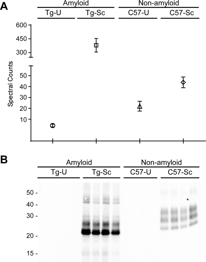
Figure 3.
Comparison of PrP in brain homogenates. (A) A spectral count comparison of PrP between sample groups shows that total PrP concentrations are lowest in the uninfected Tg44 brains (Tg-U) and highest in the RML infected Tg44 brains (Tg-Sc). Data shown are the mean and SEM of four individual mouse brains per group. (B) Brain homogenates were subjected to treatment with proteinase K and then probed with 6D11, showing that PrPSc is present only in the prion-infected samples. C57-U = uninfected C57BL/10 mice; C57-Sc = RML infected C57BL/10 mice.

