
Fig. 4.
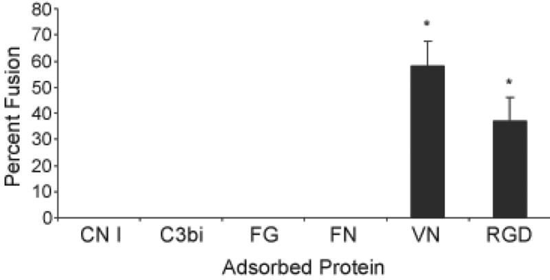
Macrophage fusion protein- or RGD-adsorbed polystyrene. Proteins or RGD peptide were adsorbed at a concentration of 25 μg per ml. Monocytes were plated and cultured for 7 days as described in Methods with the IL-4-induction of macrophage fusion on day 3. Following May-Grünwald/Giemsa staining, percent macrophage fusion was determined. Results represent mean % fusion ± SEM, n = 3 different monocyte donors. *Significantly different from other adsorbed proteins (P<0.05).