
Figure 6.
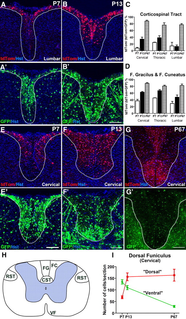
The corticospinal tract is populated mainly by dorsally derived OL lineage cells in adulthood. A, A′, There are very few OL lineage cells in the CST at P7 in the lumbar cord, and almost none of these are dorsally derived (red). B, B′, At P13, the CST at lumbar level still has very few dorsally derived OL lineage cells (red) (B), but contains many more ventrally derived (green) cells (B′). This situation reverses by P67, when the entire dorsal funiculus (DF), especially the CST, is populated mainly by dorsally derived OLPs and OLs as revealed by cell counts (C, D). The situation at thoracic and cervical spinal levels is very similar. E, E′, At P7, a majority of cells in the DF and especially the CST are ventrally derived (green). By P13, the numbers of dorsally derived (red) cells increases in the DF (F), but the CST is still mainly populated by dorsally derived cells (green, F′). The proportions of tdTom+ (dorsally derived) cells in the CST, fasciculus gracilis, and fasciculus cuneatus increase steadily from P7 to adulthood at all spinal levels (C, D). Three sections per spinal level were counted for each animal: P7 (n = 2 mice) (E, E′), P13 (n = 3) (F, F′), and P67 (n = 3) (G, G′). H, Drawing of the adult spinal cord showing the tracts and regions examined (VF, ventral funiculus). I, The absolute numbers of GFP+ (ventrally derived) cells/section in the dorsal funiculus (cervical level) decrease dramatically between P7 and P67, while tdTom+ (dorsally derived) cells increase in number during the same time frame. The images in A and B are of the lumbar spinal cord; all other images are of the cervical cord. Scale bars, 100 μm.