
Figure 1.
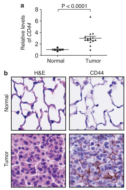
CD44 expression is increased in Kras-dependent lung adenocarcinoma. (a) The relative mRNA levels of CD44 in individual normal mouse lung tissue and KrasG12D-induced tumor samples were compared by quantitative reverse transcriptase-PCR. P values were calculated using a two-tailed Student's t test. TATA-binding protein (TBP) served as a loading control. Primer sequences used were: CD44 forward: 5′-TATGACACATATTGCTTCAATGC-3′, CD44 reverse: 5′-GTGTACCATCACGGTTGACA-3′, TBP forward: 5′-TGCACAGGAGCCAAGAGTGA-3′; TBP reverse: 5′-AGCTGGGAAGCCCAACTTCT-3′. (b) Images (original magnification, × 40) of immunohistochemistry staining of CD44 in normal mouse lung and Kras-induced tumor. Hematoxylin and eosin (H&E) stains on the left panels show normal and tumor lung epithelial cells.