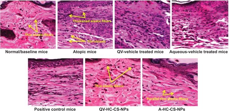
Figure 9.
Histological photomicrographs of AD-like skin lesions of NC/Nga mice treated with NP-based formulations compared to baseline mice and other treated groups.
Notes: The wavy fibers, which were stained black with VVG, represent elastic fibers. Photomicrographs were imaged under 400× magnification.
Abbreviations: A, aqueous-based; AD, atopic dermatitis; CS, chitosan; HC, hydrocortisone; NP, nanoparticle; QV-, QV-cream; VVG, Verhoeff–Van Gieson staining.

