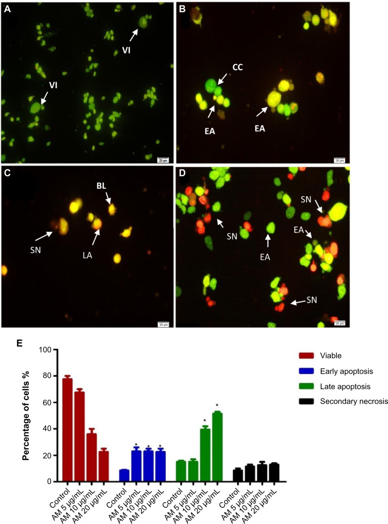
Figure 2.
Fluorescent micrographs of acridine orange and propidium iodide double-stained MDA-MB-231 cells.
Notes: (A) Untreated cells showed normal structure without prominent apoptosis and necrosis. (B) EA features were seen after treatment with 5 μg/mL of AM, with intercalated acridine orange (bright green) showing among the fragmented DNA. (C) Blebbing and orange color, representing the hallmark of late apoptosis, were noticed after 10 μg/mL AM treatment. (D) SN (bright red color) was visible after treatment with 20 μg/mL of AM. (E) Percentages of viable, early apoptotic, late apoptotic, and secondary necrotic cells after AM treatment. MDA-MB-231 cell death via apoptosis increased significantly (*P<0.05) in a concentration-dependent manner. However, no significant (P>0.05) difference was observed in the cell count of necrosis.
Abbreviations: AM, α-mangostin; BL, blebbing of the cell membrane; CC, chromatin condensation; EA, early apoptosis; LA, late apoptosis; SN, secondary necrosis; VI, viable cells.

