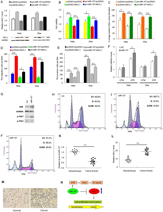
Figure 3. MCL1 rescues miR-107-induced cellular phenotypes in cervical cancer cells.
(A), cells were co-transfected with the pcDNA3/MCL1 vector, which did not contain the 3′-UTR of MCL1, with or without pri-miR-107 vector. (B–D), at 48 h after transfection, the MCL1 protein level was measured by Western blotting. MTT assay (B), colony formation assay (C), transwell assays without Matrigel (D), or Transwell assays with Matrigel (E) were used to evaluate the cell viability, growth capacity, and potential for cell migration and invasion, respectively. (F) ATR and ATM mRNA levels were monitored by qRT-PC R at the indicated time points after miR-107transfection. GADPH was used for normalization. Data represent means ± standard error (SE) from three independent experiments. (G) Western blot assay revealed that after miR-107 overexpression, ATR was upregulated, then phosphorylation of Chk1 was increased, but phosphorylation of Chk2 was no difference. (H, I and J) Cell cycle distribution was evaluated in cells transfected with negative control, miR-107 or MCL1 by propidium iodide staining and flow cytometry 24 h after transfection. N and O, the relative expression of miR-107 (K) and MCL1 (L) in the 15 pairs, including cervical cancer tissues and matched normal tissues, was determined using qRT-PCR. M, expression of MCL1 in cervical cancer tissue and normal tissues by immunohistochemistry (n = 12). N, schematic representing miR-107-mediated regulation of MCL1 expression during cancer progression. Model of modified ATR/Chk1 pathway, in which miR-107 and MCL1 are involved. Data represent means ±SE from three independent experiments. Significance was determined by Student's t-test: *p<0.05, **p<0.005.

