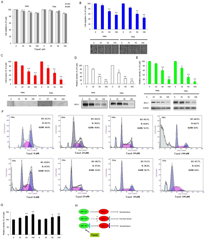
Figure 4. Effects of taxol on the viability, migration, and invasion in cervical cancer cells.
(A)The chemical structure of taxol. (B) HeLa cells and SiHa cells were treated with increasing concentrations of taxol for 24 and 48 h. Cell viability was determined by MTT assay. The plot presents relative cell viability compared to untreated (0 µM) cells. HeLa and SiHa cells were treated with the indicated concentrations of taxol for 48 h. Subsequently, the migratory (C) and invasive (D) ability of cells after each treatment were determined, as described in material and methods. The bottom plots were the relative cell numbers comparing to that in untreated (0 µM) cells. Cells were treated with the indicated concentrations of taxol for 48 h. (E) the conditioned medium from each treatment was collected, and MCL1 activity was determined by casein zymography. (F) Cell lysate was applied to determine the protein levels of MCL1 by Western blotting. GAPDH was used as the internal control. (G) Apoptosis assay showed that taxol suppressed cell apoptosis and G1 arrest. (H) the relative expression of miR-107, (I) the working model of taxol regulates expression of MCL1 by down-regulating miR-107. Bars show the value as mean ±S.E. from three independent experiments. *, P<0.05; **, P<0.005, compared with the untreated cells.

