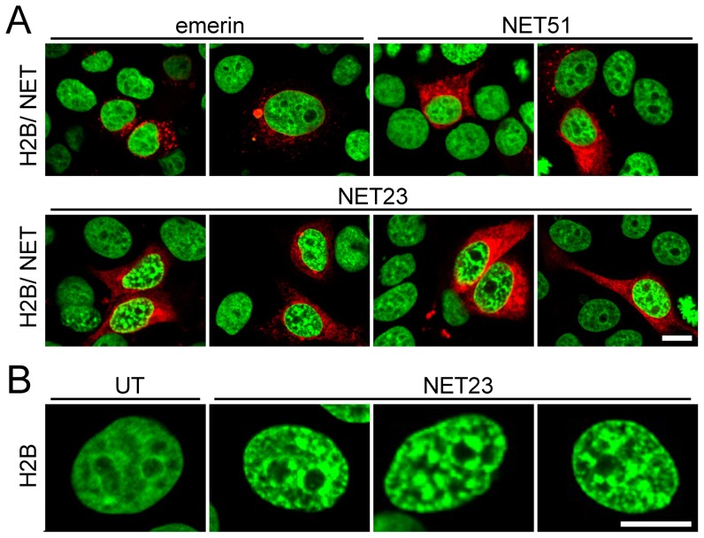
Figure 1. A screen for NETs that alter chromatin compaction.
(A) 72 h post-transfection HeLa cells have no gross changes in distribution of H2B-GFP (green) when most NETs fused to mRFP (red) are exogenously expressed (e.g. emerin and NET51, upper panels). However, cells transfected with NET23/STING (lower panels) exhibit considerable chromatin compaction. (B) Zoomed images of chromatin in untransfected (left) and NET23 transfected cells. All images were taken using identical settings and all scale bars = 10 µm.

