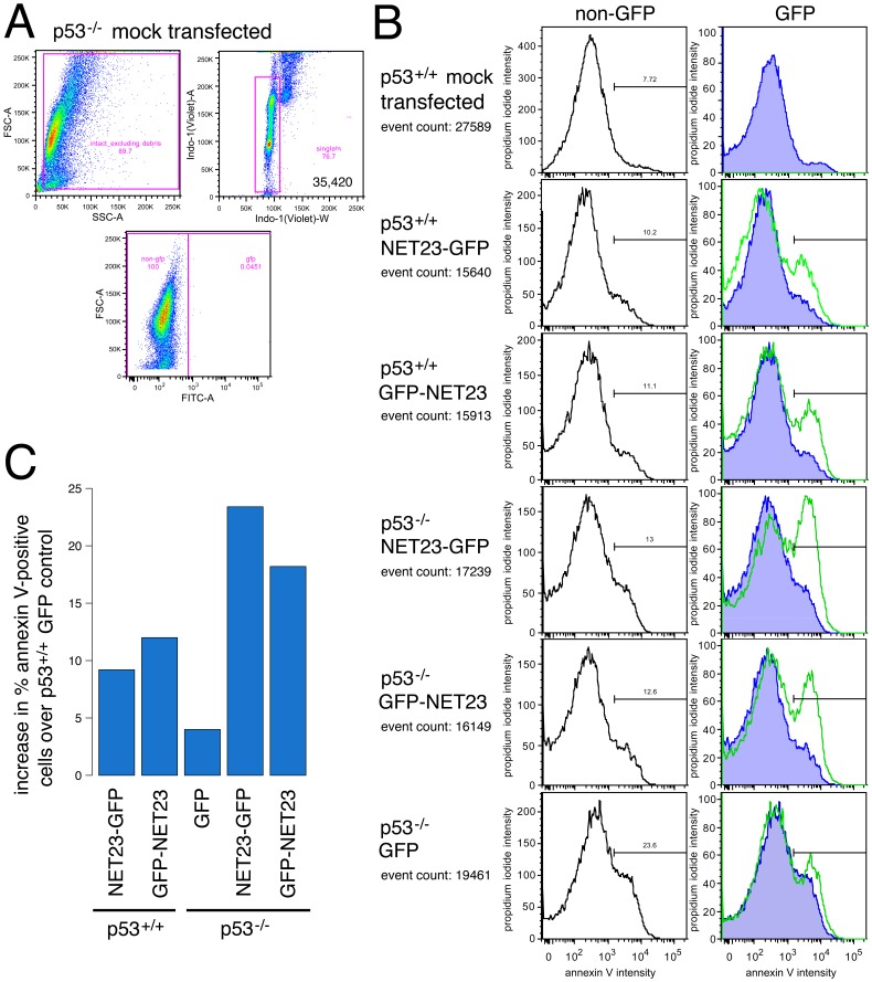
Figure 7. NET23/STING promotes apoptosis.
(A) Gating strategy used for cells in B using forward and side scatter profiles to exclude debris followed by DNA content to determine intact singlet cells. The transfected population (expressing GFP) was identified by subsequently gating singlet cellular material on forward scatter versus GFP intensity. All cells in this experiment were analyzed at 44 h post-transfection. (B) The cells used to determine the gates were also stained for propidium iodide (PI; y-axis) and annexin V (x-axis). The traces in the left panels show the untransfected cells in the population and those in the right panels show the cells with GFP signal. The right-most green peak delineates cells with an annexin V signal of sufficient intensity to indicate cells undergoing apoptosis. As expected, for the mock-transfected culture essentially no GFP positive cells were identified and very few apoptosing cells could be observed. Expression of NET23/STING consistently increased the apoptosing population regardless of whether the tag was on the N-terminus (GFP-NET23) or the C-terminus (NET23-GFP) and the effect of NET23/STING did not require function of the master regulator p53 as apoptosis was induced in both wild-type (p53+/+) and p53 knockout (p53−/−) cells. Nonetheless, it is notable that the responses were very similar between the two NET23/STING constructs in the wild-type cells while the N-terminal tag showed a lagging apoptotic response in the p53 knockout cells. (C) The percentage of annexin V-positive cells is plotted after correction to subtract the number in the GFP control with the wild-type (p53+/+) cells. This is used as the correction for both cell lines to better indicate the effect of the p53 knockout itself on apoptosis induction.

