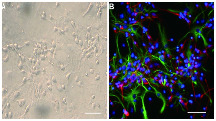
Figure 1. Identification of cerebral cortical cells and neural stem cells.
The cerebral cortical cells were cultured with Neurobasal medium containing 2% B27 for 5 d; the spindly neurites grew out of the cell bodies and were observed as three-dimensional structures (A). Immunofluorescence staining showing nuclei stained blue with Hoechst33258, immunopositive neurons stained red with β-TubIII, and immunopositive astrocytes stained green with GFAP (B). Scale bar = 200 µm.

