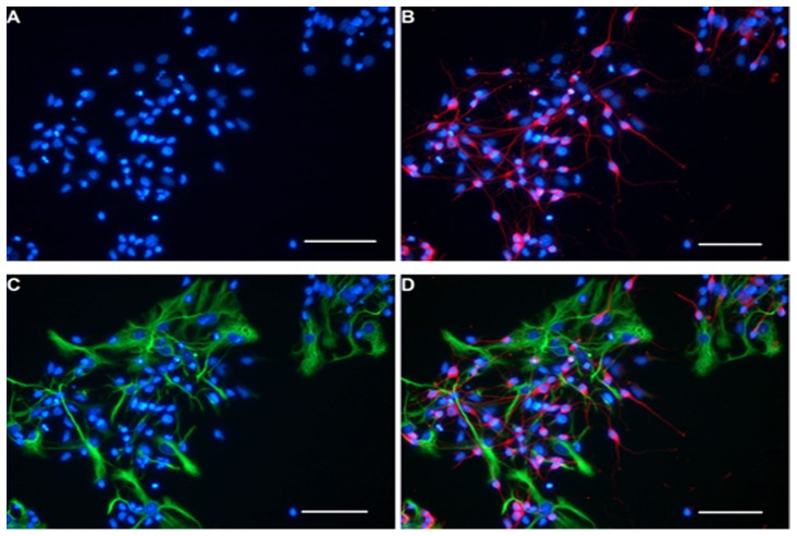
Figure 3. Immunofluorescence identification of differentiated NSCs.
Neurons and astrocytes derived from NSCs were immunoreactive with anti-β-TubIII and anti-GFAP respectively. All of the nuclei were stained blue with Hoechst33258 (A), β-TubIII+ neurons were stained red (B), and GFAP+ astrocytes were stained green (C). Immunostaining showed that the marker β-tubIII and GFAP never co-localization at the same field (D). Scale bar = 100 µm.

