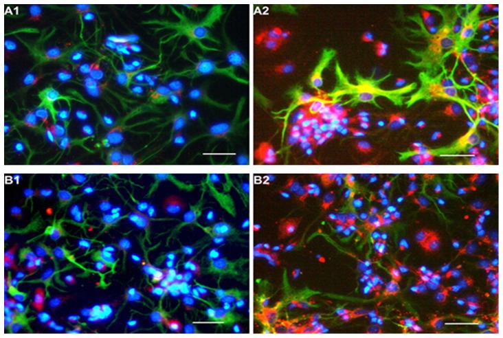
Figure 4. Immunofluorescence identification of VEGF and BDNF in cerebral cortical cells.
Astrocytes (GFAP+) were stained green, nuclei were stained blue with Hoechst33258, both VEGF+ and BDNF+ cells were stained red. A1 and B1 represent the cells cultured with NCM, B1 and B2 represent the cells cultured with 4% HCM. Expression of VEGF and BDNF was observed in some of the astrocytes (yellow staining in the astrocytes). Scale bar = 100 µm.

