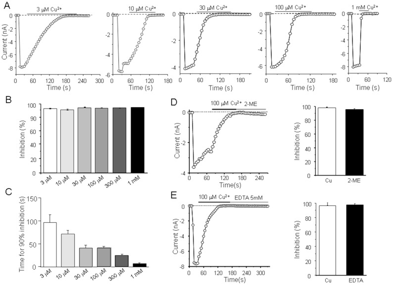
Figure 1. TRPM2 open channels inactivated by extracellular Cu2+.
(A) Representative recordings of the inward currents evoked by 500 µM ADPR at −80 mV, using a 500 ms voltage ramp of −100 mV to +100 mV applied every 5 s, before and after exposure to the indicated Cu2+ concentrations. The dotted lines indicate zero currents. (B–C) Summary of the percentage inhibition (B) and time required for inward current amplitude reached 90% inhibition after Cu2+ exposure (C). (D) Left panel, the ADPR-induced inward currents when fully inhibited by 100 µM Cu2+ were not reversed after treating with 20 µM 2-ME; Right panel, summary of the current recovery during exposure to 2-ME. (E) Left panel, the ADPR-induced inward currents when fully inhibited by 100 µM Cu2+ were not reversed after treating with 5 mM EDTA; Right panel, summary of the current recovery during exposure to EDTA. Residual current expressed as the percentage of the currents immediately before exposure to Cu2+ is 3.3±1.7% after inactivation by Cu2+, which returned to 2.6±0.9% after washing with EDTA. In 2-ME group, residual current changed from 1.8±0.5% to 2.0±0.7%. The number of cells examined in each case is 4–6.

