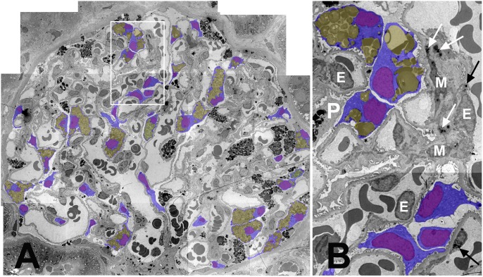
Figure 1. Mosaicism of podocyte Fabry phenotype in a glomerulus from a female patient with Fabry disease.
(A) Montage image of a glomerulus (∼3,000×). Podocyte bodies with visible nuclei are colored blue, podocyte nuclei purple, and GL-3 inclusions yellow. The white rectangle is magnified in B. (B) Magnified view of three podocyte profiles without (at the bottom) and three other podocyte profiles with GL-3 inclusions (on the top). Arrows show GL-3 inclusions in mesangial (M) cells (black) and endothelial (E) cells. P is a podocyte profile with no visible nucleus on this section.

