
Figure 2. Loss of clock plasmid in the intracellular fraction and estimated mycobacterial replication and death rates.
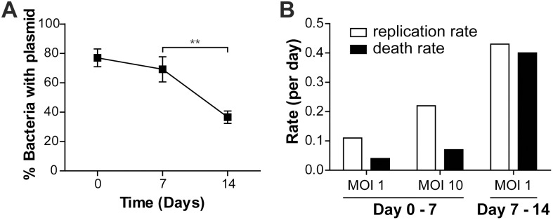
(A) CFU counts from cell lysates during MOI 1 infection on kanamycin-containing plates normalized to total CFU counts on plates without kanamycin. Differences in percentage of bacteria containing the clock plasmid was analyzed using 1-way ANOVA and Tukey’s post-hoc test. n = 8–11. **p<0.01. (B) Estimated replication and death rates (per day) for intracellular Mtb were calculated from clock plasmid CFU data. Rates for MOI 10 infection between day 7 and 14 could not be determined due to extensive cell death.