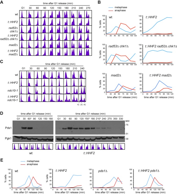
Figure 1.

Histone depletion activates the SAC. (A, B) Metaphase arrest by histone loss is Mad2-dependent and Rad53 and Chk1-independent. Cell cycle progression analysis by flow cytometry (A) and nuclear and spindle morphologies by DAPI and immunofluorescence against tubulin (B) of wild-type, t::HHF2, sml1Δ rad53Δ chk1Δ, sml1Δ t::HHF2 rad53Δ chk1Δ, mad2Δ and t::HHF2 mad2Δ cells synchronized in G1 and released into fresh medium under conditions of histone depletion. sml1Δ did not affect cell cycle progression of wild type and t::HHF2 (data not shown). (C) A functional kinetochore is required for SAC activation in t::HHF2. DNA content was analyzed by flow cytometry of wild-type, ndc10-1, t::HHF2 and t::HHF2 ndc10-1 cells synchronized in G1 at permissive temperature (26°C) and released into fresh medium at restrictive temperature (37°C) under conditions of histone depletion. (D) Securine/Pds1 remains undegraded during SAC activation in t::HHF2 cells. Pds1 accumulation was determined by western analysis of wild-type and t::HHF2 cells synchronized in G1 and released into fresh medium for one cell cycle under conditions of histone depletion. Asterisks show the time at which α-factor was added to prevent cells from re-entering a new mitosis. MW, molecular weight marker. Cell cycle progression by DNA content analysis is shown below. (E) Securine/Pds1 is required for SAC activation in t::HHF2 cells. Cell cycle progression was analyzed by immunofluorescence against tubulin (spindle morphology) for wild-type, pds1Δ, t::HHF2 and t::HHF2 pds1Δ cells synchronized in G1 and released into fresh medium at 23°C under conditions of histone depletion.
