
Figure 5.
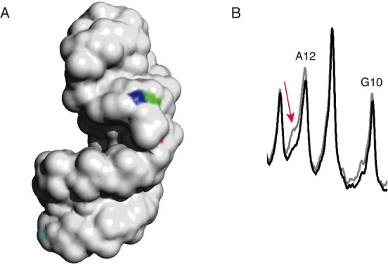
The 2″- and 3′-hydrogen atoms of residue G10 are highly solvent exposed. (A) Solvent-exposed surface of the SRL. The 2″-hydrogen atom of residue G10 is colored blue; the 3′-hydrogen atom of residue G10 is colored green. The 2″-hydrogen atoms of all other residues are colored cyan, and the 3′-hydrogens are colored orange. (The 2″- and 3′-hydrogens of residues other than G10 are mostly not visible in this image because their solvent accessibilities are very low.) (B) Overlaid scans of gel lanes in which were separated cleavage products of SRL containing all natural nucleotides (gray), and SRL in which [2″-2H]-guanosine had been incorporated (black). The SRL was radiolabeled at the 5′ end. While almost no difference in intensity is seen for the band assigned to cleavage at residue G10, the shoulder on the band assigned to cleavage at residue A12 decreases in intensity upon 2″-deuteration of guanine (red arrow). The mobility of this product is consistent with that of the 3′-deoxy-2′-keto-terminated strand that would result from abstraction of a 2″-ribose hydrogen atom from residue G10 (see text).