
Figure 11.
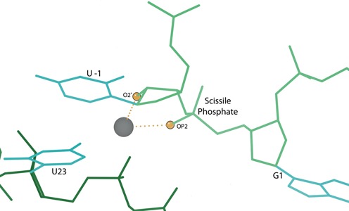
The position of G1, modeled U -1 and the scissile phosphate in 3NKB, based on the active site of the 2OEU hammerhead ribozyme structure (22). Modeled U -1 stacks with U23 and the dotted lines show the metal ion coordination by OP2 (pro-Rp) of the scissile phosphate and O2′ of U -1 (highlighted as balls).