
Figure 3.
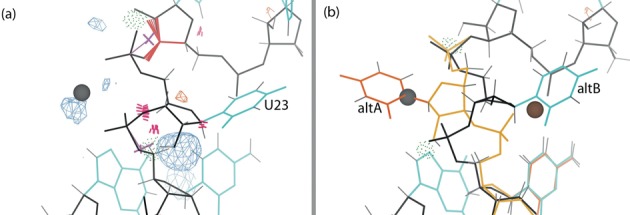
U23 and Mg2+ of the cleaved structure with Fo-Fc difference density at 3.5σ contour level. Here and in later figures, the blue and orange mesh correspond to positive and negative difference density respectively, hydrogen bonds are shown as green dotted pillows, steric clashes as hotpink spikes, ribose pucker outliers as magenta crosses, and bond-length or bond-angle outliers as springs or fans (blue if too small, red if too large). (a) The base and backbone of U23 in the original structure (1CX0), with the modeling errors highlighted. (b) Alternate conformations of U23 in the rebuilt structure (4PR6), with the backbone (gold), base (orange) and metal ion (brown) for the new conformation. All the modeling errors are corrected, and the difference density peaks have disappeared.