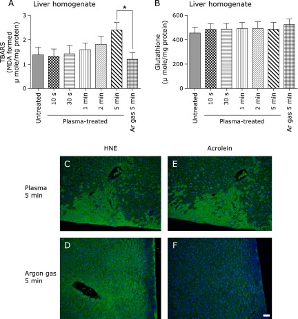
Fig. 3.
Determination of TBARS and Glutathione reduced form (GSH) in rat liver homogenate and detection of HNE-modified proteins and acrolein-modified proteins by immunohistochemical analyses in rat liver. (A) TBARS in rat liver homogenates were significantly elevated after NEAPP irradiation for 5 min. (B) GSH in rat liver homogenate was unchanged after NEAPP treatment. Representative merged images of immunohistochemical staining for HNE-modified protein (C, D) and acrolein-modified proteins (E, F) are shown (scale bar, 50 µm). HNE and acrolein staining disclosed a depth-dependent spread in the hepatic parenchyma only after 5 min of NEAPP irradiation (C, E). There was no specific immunostaining in argon gas treated samples with a background diffuse staining in HNE-modified proteins (D, F). Data are expressed as means ± SEM (n = 5; *p<0.05 vs argon gas for 5 min).

