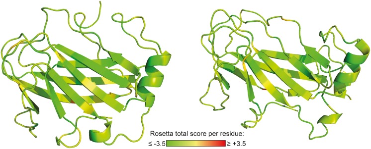
Figure 4. Structural models for laminin γ1 L4.
Shown are the two best-scoring models generated by comparative modeling based on 13 structural homologues that have been identified by fold recognition. The residues are colored according to their Rosetta total score. Scores below zero (yellow-green color) indicate energetically favorable conformations.

