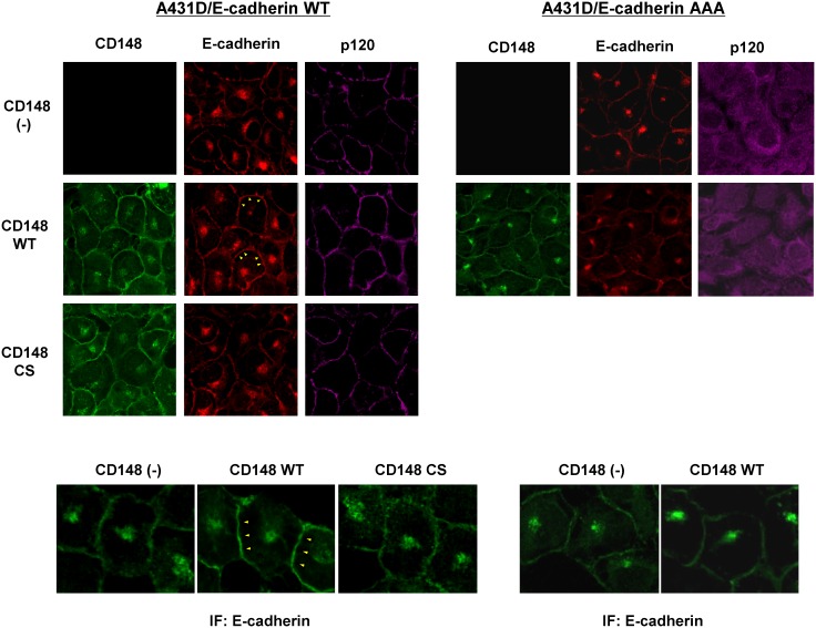
Figure 2. Effects of CD148 in E-cadherin distribution.
Immunofluorescence localization of CD148 (green), E-cadherin (red), and p120 (purple) were examined in CD148 WT or CS-introduced A431D/E-cadherin WT (left panels) and A431D/E-cadherin 764AAA (right panels) cells and compared with CD148-negative cells. Lower panels show a higher magnification of E-cadherin immunofluorescence. Wild-type E-cadherin is more broadly distributed at cell junctions in CD148 WT-introduced cells (arrowheads in left panels), while the distribution of p120-uncoupled E-cadherin is unaltered by CD148 WT introduction (right panels).

