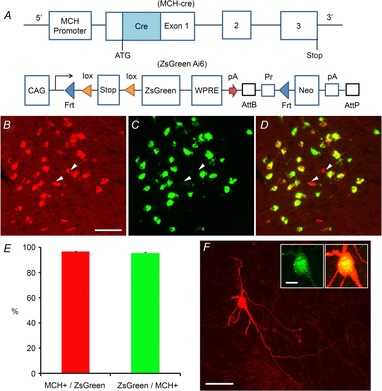
Figure 2. Generation of the pMCH-cre/ZsGreen mouse and characterization of ZsGreen-expressing MCH+ cells in the lateral hypothalamus.

A, schematic diagram showing the transgenic regions of the MCH-cre and ZsGreen (Ai6) reporter mouse lines. The pMCH-cre mouse (Kong et al. 2010) is crossed with the Ai6 mouse (Madisen et al. 2010) to generate the pMCH-cre/ZsGreen mouse. In the ZsGreen reporter mouse, the Rosa26 locus is modified by targeted insertion of a construct containing the strong and ubiquitously expressed CAG promoter, followed by a loxP-flanked (‘floxed’) stop cassette-controlled fluorescent marker gene (ZsGreen). The construct uses the woodchuck hepatitis virus post-transcriptional regulatory element (WPRE), and a pair of PhiC31 recognition sites (AttB/AttP) through which the Neo marker can be deleted from the reporter line. The FRT sites allow a Flp recombinase-mediated replacement strategy (‘Flp-in’) to swap other genes into the same locus at high efficiency. B–D, ZsGreen-expressing cells are MCH+ neurons in the lateral hypothalamus of the pMCH-cre/ZsGreen mouse. B, C and D are example images of ZsGreen expression in the lateral hypothalamus, immunostaining of MCH in the same section, and the merge of B and C, respectively. The arrowheads point to examples of a few MCH-immunopositive cells which do not express ZsGreen. Scale bar: 50 μm. E, summary of immunochemical data quantification obtained from 4 pMCH-cre/ZsGreen mice. F, typical morphology of single ZsGreen+/MCH+ neurons, revealed by post hoc biocytin staining. The insets show that this example cell expresses MCH. Scales: 50 and 10 μm.
