
Figure 8. Schematic diagram of how histamine may regulate the MCH system during sleep and wakefulness.
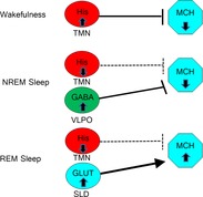
During wakefulness, histaminergic neurons in the tuberomammillary nucleus (TMN) silence MCH neurons. During NREM sleep, histaminergic neurons are inactive and do not affect MCH neurons, but NREM-active GABAergic neurons in the ventrolateral preoptic area (VLPO) keep MCH neurons silent. During REM sleep, wake-active and NREM-active neurons are silent and MCH neurons are activated by REM-active glutamatergic neurons in the sublaterodorsal nucleus (SLD).