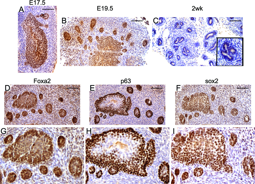
Figure 2. SOX2 expression during murine embryonic prostate development.
Nuclear SOX2 staining was detected in the prostatic buds at E17.5 and E19.5 (A&B). At 2 weeks after birth, the nuclear staining of SOX2 was hardly detected in prostate (C, inset is a higher magnification picture). The prostatic buds co-expressed Foxa2 (D), p63 (E), and SOX2 (F). G, H, and I are higher magnification picture of D, E, and F, respectively. Scale bars represent 25 µm.

