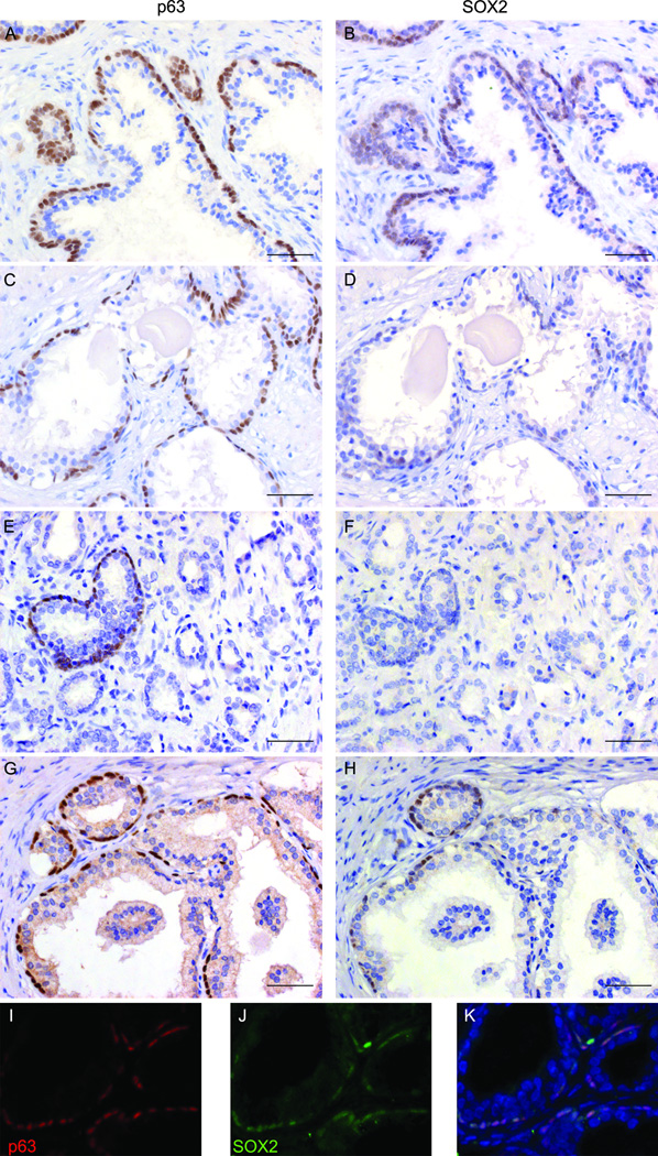
Figure 6. Expression of SOX2 in human BPH and primary prostate adenocarcinoma specimens.
Immunohistochemical staining of SOX2 or p63 was performed on serial sections derived from BPH (A–D) or PCa (E–H) tissues. Panels A&B represented the 26 of 30 BPH cases that displayed positive SOX2 staining in basal cell layer; panels C&D represented the 4 of 30 BPH cases that showed little or no SOX2 staining but were still positive for p63. Panels E&F represented the 22 of 24 primary PCa cases that lost SOX2 expression. Some benign areas in these PCa specimens were positive for p63 but negative for SOX2 staining. Panels G&H represented cancer-adjacent normal areas in the 2 of 24 PCa cases where both p63 and SOX2 were expressed. Panels I-K are images from dual immunofluorescence staining performed on sections derived from BPH specimens. SOX2 (in green) was co-expressed with basal cell marker-p63 (in red). DAPI was used for counter-staining. Scale bars represent 25 µm.

