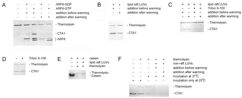
Figure 1.
ARF6- or lipid raft-induced alterations to the structure of CTA1.
A. As indicated, equimolar amounts of ARF6/GDP or ARF6/GTP were added to either folded CTA1 before warming to 37°C or to CTA1 disordered by warming for 30 min at 37°C. Following a total of 1 h at 37°C, toxin samples were placed on ice and exposed to the protease thermolysin for 1 h at 4°C. Proteins were detected by sodium dodecyl sulfate polyacrylamide gel electrophoresis (SDS-PAGE) with Coomassie staining. CTA1 was initially present in all samples. The addition of GTP induces a conformational change in ARF6 that renders it susceptible to nicking by thermolysin.
B. Lipid raft LUVs were added to either folded CTA1 before warming to 37°C or to CTA1 disordered by warming for 30 min at 37°C. Following a total of 1 h at 37°C, toxin samples were placed on ice and exposed to thermolysin for 1 h at 4°C. Proteins were detected by SDS-PAGE with Coomassie staining.
C. Lipid raft LUVs in the absence or presence of 1% Triton X-100 were added to either folded CTA1 before warming to 37°C or to CTA1 disordered by warming for 30 min at 37°C. Following a total of 1 h at 37°C, toxin samples were placed on ice and exposed to thermolysin for 1 h at 4°C. Proteins were detected by SDS-PAGE with Coomassie staining.
D. CTA1 was incubated at 25°C for 1 h in the presence or absence of 1% Triton X-100. Toxin samples were then placed on ice and exposed to thermolysin for 1 h at 4°C. Proteins were detected by SDS-PAGE with Coomassie staining.
E. Samples of α-casein were incubated for 1 h at 4°C with thermolysin in the absence or presence of lipid raft LUVs. Proteins were then detected by SDS-PAGE with Coomassie staining.
F. Non-raft LUVs mimicking the charge and fluidity of the plasma membrane were added to either folded CTA1 before warming to 37°C or to CTA1 disordered by warming for 30 min at 37°C. Additional CTA1 samples were incubated at 37°C or 25°C in the absence of LUVs. Following a total of 1 h at the indicated temperature, toxin samples were placed on ice and exposed to thermolysin for 1 h at 4°C. Proteins were detected by SDS-PAGE with Coomassie staining.

