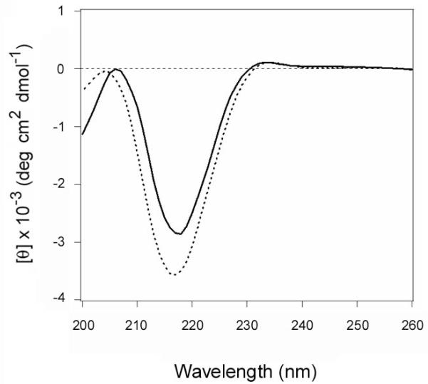
Figure 9.
Structures of reduced CTA1 from the CTA1/CTA2 heterodimer and the recombinant His-tagged protein.
A purified CTA1/CTA2 heterodimer was treated with 30 mM GSH and subjected to dialysis in 30 mM GSH buffer with a 3500 MWCO filter. Far-UV CD was then used to record the spectrum of the retained CTA1 subunit (solid line). A far-UV CD spectrum was also recorded for CTA1-His6 in the presence of 30 mM GSH (dashed line). Both measurements used 50 μg of toxin in 220 μL of 10 mM sodium borate buffer (pH 7.0) containing 100 mM NaCl. Measurements were recorded with a Jasco 810 spectrofluoropolarimeter at 18°C, and the spectra were averaged from three scans.

