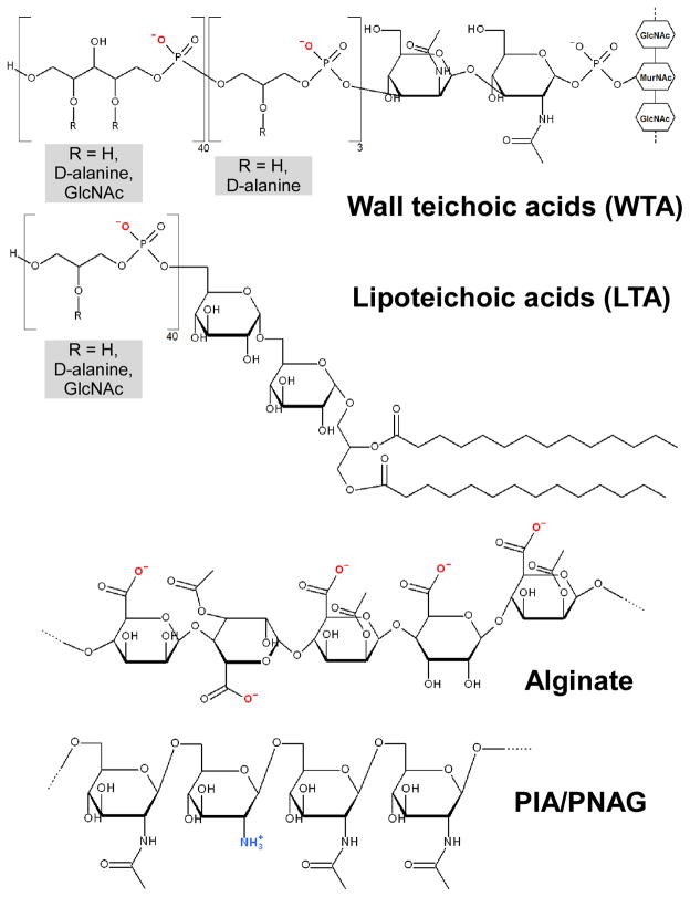
Fig. 5. Examples of bacterial surface polymers.
Wall teichoic acids (WTA) and lipoteichoic acids (LTA) occur in Gram-positive bacteria. The structures found in S. aureus are shown. WTA is linked via a phosphodiester bond to an N-acetyl muramic acid residue in peptidoglycan. The 2 OH-group of N-acetylmannosamine is phosphodiester linked to three units of (D-alanylated) glycerol-phosphates, to which ~ 40 units of ribitol phosphates (substituted with D-alanine and/or GlcNAc) are linked. In LTA, a diacylglycerol moiety, which is linked to a β-1,6-connected diglucosyl part, forms the membrane anchor. Approximately 40 units of glycerol phosphate are linked to the 6-hydroxyl group of the second glucosyl part. Position 2 of the glycerol phosphate residues is in part substituted either with D-alanine or D-alanine ester and glycosyl (mostly GlcNAc) residues. GlcNAc, N-acetyl-glucosamine. Alginate (of P. aeruginosa) is composed of guluronic acid (GulUA) and mannuronic acid (ManUA) monomers. PIA/PNAG (S. aureus, S. epidermidis, other staphylococci and some other bacteria) is a homopolymer of partially de-acetylated N-acetyl-glucosamine units in β-1-6 linkage.

