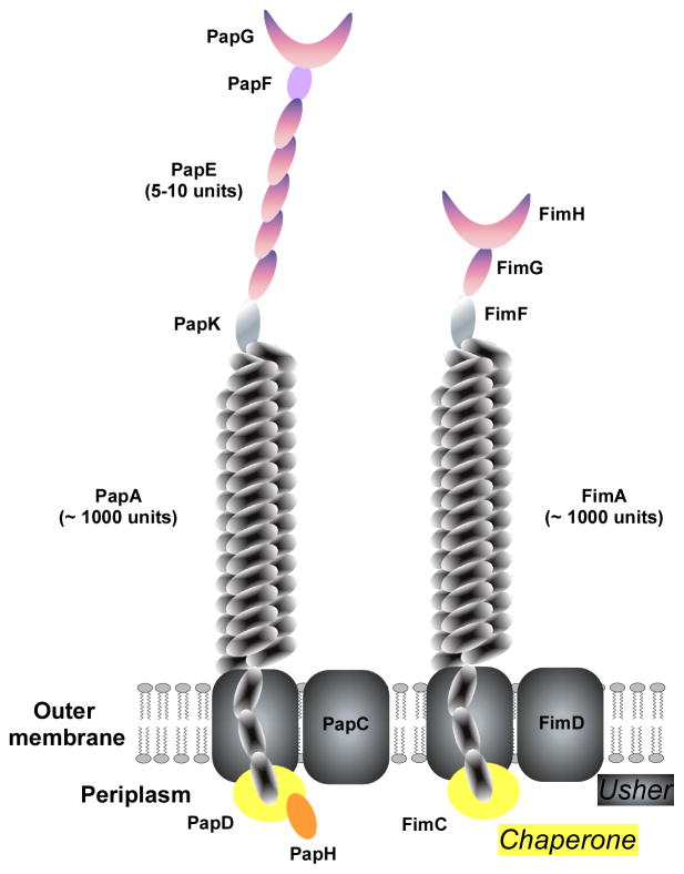
Fig. 8. Structure of pili of the chaperon-usher-secreted type (P and type 1 pili).
Type I pili of Gram-negative bacteria are secreted and linked to the outer membrane by the chaperone-usher pathway. Type 1 and P pili are the most important examples, shown here. PapC and FimC represent the ushers, while PapD/PapH and FimC, respectively, are the chaperones. The main part of the pilus is made of and linked to the outer membrane by the PapA and FimA protein units, of which ~ 1000 assemble. PapG and FimH are the adhesin molecules situated on the tip of the pilus. Between the PapA or FimA multimers and the PapG/FimH tips, type 1 and P pili differ somewhat in structure, with the type 1 pilus having additional subunits (see graph).

