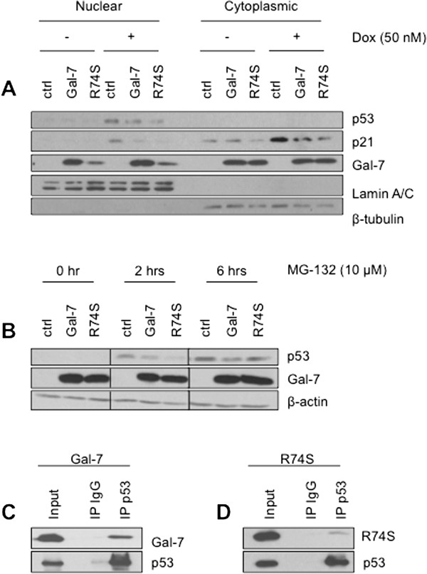
Figure 7.

Decreased of p53 nuclear translocation through proteasomal degradation induced by cytoplasmic gal-7. (A) Cytosolic and nuclear fractions were purified from control (srα) cells and MCF-7 cells expressing wild type or mutated gal-7 after doxorubicin treatment. Expression of p53, p21 and gal-7 was measured in both fractions by Western blotting. β-tubulin and lamin A/C are shown as positive cytoplasmic and nuclear expression controls, respectively. (B) Cells were treated with 10 μM MG-132 for 0, 2, and 6 hrs. Total cellular extracts were subjected to Western blot analysis for p53, gal-7 and β-actin. Immunoprecipitation (IP) experiments showing p53 interaction with (C) wild type and (D) R74S mutant of gal-7 in MCF-7 cells. Whole-cell lysates were made from MCF-7 cells transfected with a construct expressing p53. Lysates were immunoprecipitated with anti-p53 or a control IgG antibody, and immunoblot analysis was performed with anti-gal-7.
