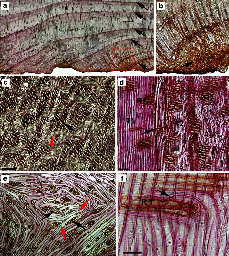
Fig. 2.
Microscopic sections of the overgrown tissue. a Transverse section of regenerative tissue with distinctive layers of annual rings of wood. b Tissue sector marked on a under higher microscopic magnification; the callus-like parenchymal zone with unorganized cells formed at the beginning of tissue regeneration (arrow). c Radial and e tangential sections of overgrown tissues in the region of the circular tracheary elements’ differentiation (red arrow) around the ray-like structures (black arrows). d Radial section of stump tissue, below the cut surface, formed before and after stem cutting. T and R, tracheids and rays formed before (1) and (2) after cutting; arrow, boundary between xylem formed before and after stem cutting. f Radial section of stump xylem below the cut surface, formed after stem cutting, under higher microscopic magnification. R, xylem ray; arrow, tip of tracheid. Scale bars = 350 μm (a), 50 μm (b), 150 μm (c, d), 100 μm (e), 60 μm (f)

