
Fig. 6.
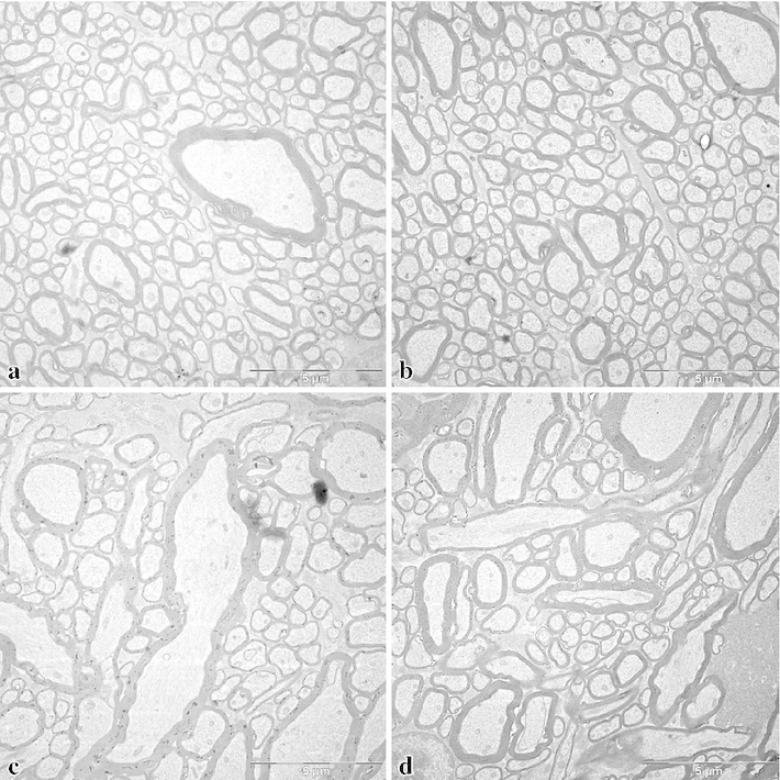
Electron micrographs from the region of the superior longitudinal fascicle of the macaque brain (M. mulatta). a and b show regions where all fibres run orthogonally to the frontal sections, c and d show a neighbouring region with fibres running also in other directions. This region was not included in the measurements, but is shown here for illustrating different aspects of the cortical white matter. Bars 5