Figure 1.
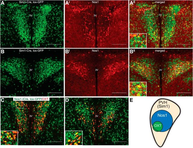
Neuronal Nos1 marks a subset of PVH neurons. A, B, IHC for NOS1 peptide (red) in the PVH of Sim1-Cre, lox-GFP reporter mice (lox-GFP, green) identifies Nos1PVH neurons as a Sim1PVH neuronal subset. C, D, OXTPVH neurons are contained within the Nos1PVH population (green), as shown by expression of OXT (red) in sections from Nos1-iCre, lox-GFP mice. E, A model of neurochemically defined cell types within the PVH. Dashed boxes indicate regions that are digitally enlarged and shown as insets. Arrowheads indicate representative overlapped cell-bodies. Scale bar, 200 μm. 3V, Third ventricle.

