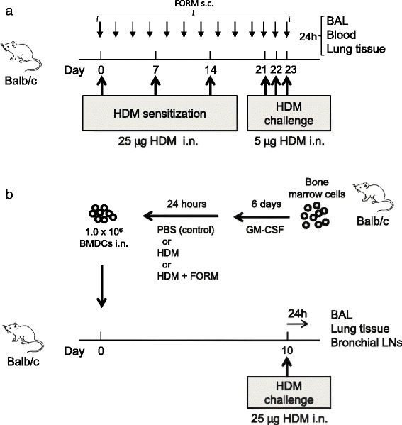
Figure 1.

Protocols for the HDM-induced airway inflammation model. (a) HDM-induced airway inflammation model. Female BALB/c mice were sensitized three times intranasally with 25 μg HDM at days 0, 7 and 14, and challenged three times intranasally with 5 μg HDM at days 21–23. Twenty-four hours after the final challenge, blood samples, BAL fluid and lung tissues were collected. BALB/c mice receiving PBS at sensitization and challenge were used as controls. (b) BM cells were harvested from BALB/c mice. DCs were generated by culturing BM cells with 10% FBS and 10 ng/ml GM-CSF in the culture medium for 7 days. On day 7, DCs were stimulated with HDM or HDM + FORM or PBS (control) for 24 hours. One million DCs were intranasally injected into BALB/c mice on the following day. After 10 days, recipient mice were challenged intranasally with 25 μg HDM. Twenty-four hours after the injection, blood samples, BAL fluid and lung tissues were collected. BALB/c mice receiving unstimulated DCs served as controls.
