
Figure 1.
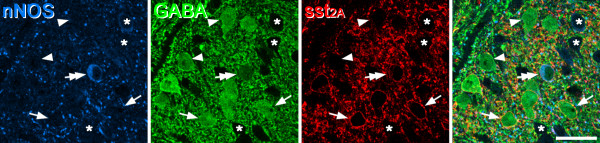
nNOS, GABA and sst2A in lamina II in the mouse. A single confocal scan through a section that had been reacted to reveal nNOS, GABA and sst2A. A nNOS+/GABA+ neuron (double arrow) is sst2A+. Several other GABA+ cells that lack nNOS are present in this field. Two of these that are sst2A+ are marked with arrows, while two that are sst2A- are indicated with arrowheads. Three nearby GABA-negative cells are labelled with asterisks. Scale bar: 20 μm.