
Figure 3.
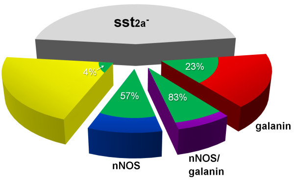
Expression of galanin and nNOS among sst2A+ neurons in the superficial dorsal horn, and their relationship with GFP in the PrP-GFP mouse. The pie chart shows the sizes of different neurochemical populations among the inhibitory interneurons in laminae I-II. We have estimated that 54% of the inhibitory interneurons in this region are sst2A-immunoreactive in a different mouse strain (C57Bl/6) [4], and the present results indicate that the proportion of these cells that contain only nNOS, only galanin or both nNOS and galanin are 17%, 31% and 13%, respectively. The proportions of each of these populations that are accounted for by GFP+ neurons in the PrP-GFP mouse are shown in green, with the corresponding percentages indicated.