
Figure 1.
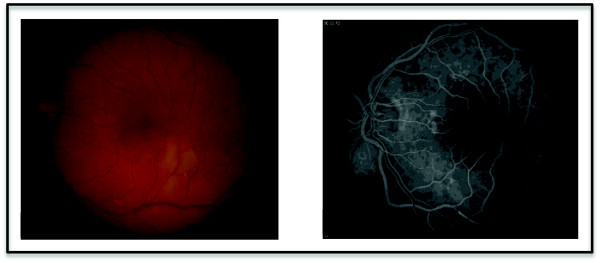
The fundus photograph demonstrates multiple Hollenhurst plaques and an area of retinal edema in the left eye. The fluoroscein angiogram highlights inferior vascular filling defects.
Official websites use .gov
A
.gov website belongs to an official
government organization in the United States.
Secure .gov websites use HTTPS
A lock (
) or https:// means you've safely
connected to the .gov website. Share sensitive
information only on official, secure websites.
The fundus photograph demonstrates multiple Hollenhurst plaques and an area of retinal edema in the left eye. The fluoroscein angiogram highlights inferior vascular filling defects.