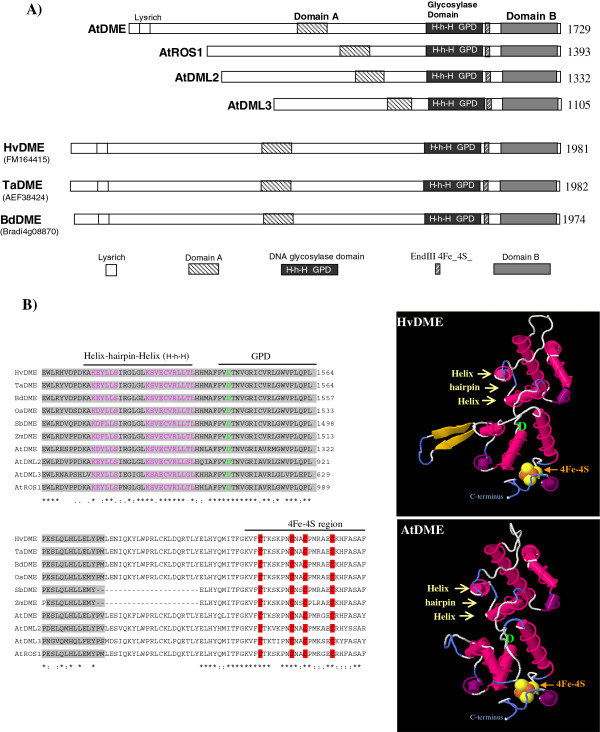
Figure 1.

Schematic view of DME-family DNA glycosylases and predicted tertiary structure of HvDME and AtDME. A) Proteins, HvDME (FM164415), TaDME (AEF38424.1), BdDME (Bradi4g08870), AtDME (NP_196076.2), AtROS1 (AAP37178.1), AtDML2 (NP_187612.5) and AtDML3 (NP_195132.3) are depicted with white rectangles. White box, lysine-rich region; black box, glycosylase domain; hatched box, A domain; grey box, B domain; grey hatched box, 4Fe-4S binding domain; H-h-H, helix-hairpin-helix; GPD, glycine-proline rich region and conserved aspartate residue. B) Left: Amino acid sequence alignment of the glycosylase domain of DME-type proteins. Right: Predicted tertiary structure of HvDME and AtDME glycosylase domains. Helix-hairpin-helix is indicated with yellow arrows, the conserved aspartate (D) is shown in green and the 4Fe-4S is shown in orange-yellow.
