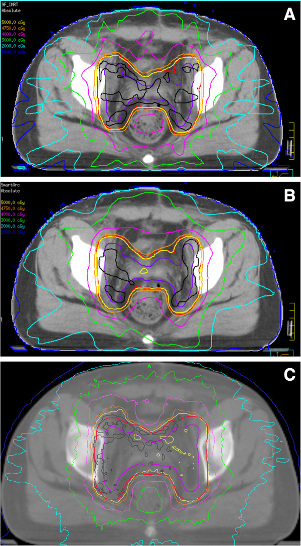
Figure 1.

Representative axial computed tomography slices showing isodose distributions. (A) IMRT. (B) VMAT-S. (C) Tomotherapy. PTV is shown in red, CTV in slate blue. Isodose lines are indicated as follows: inverse grey, 5250 cGy; yellow, 5000 cGy; orange, 4750 cGy; purple, 4000 cGy; green, 3000 cGy; sky blue, 2000 cGy; and blue 1000 cGy.
