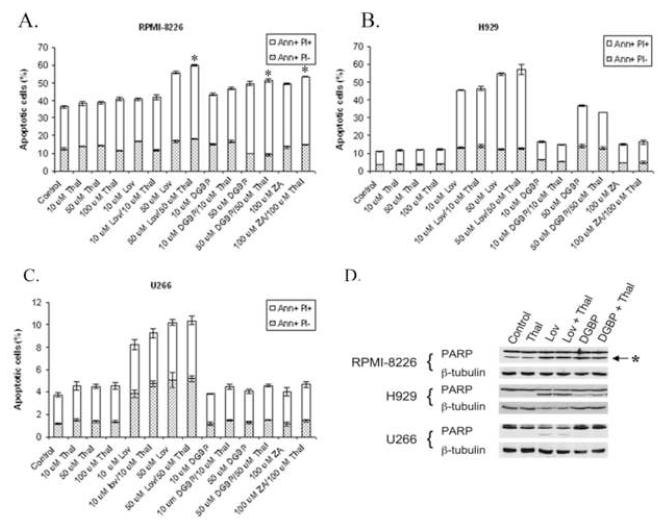
Figure 3.
The effect of thalidomide and IBP inhibitors on induction of apoptosis in myeloma cells. Annexin V, propidium iodide (PI) flow cytometric experiments were performed in RPMI-8226 (A), H929 (B), and U266 (C) cells. Cells were incubated for 48 hours. The percentages of cells in the early apoptotic (Annexin V positive, PI negative (Ann+ PI-)) and late apoptotic/necrotic (Annexin V positive, PI positive (Ann+ PI+)) fractions are shown (n =3). * denotes p<0.05 per unpaired two-tailed t-test comparing thalidomide + IBP inhibitor to IBP inhibitor alone. D) Immunoblots depicting PARP cleavage. Cells were incubated for 48 hours with combinations of thalidomide (Thal, 50 μM), lovastatin (Lov, 50 μM) or DGBP (50 μM). The “*” denotes the C-terminal PARP cleavage product. β-Tubulin is shown as a loading control.

