Figure 3.

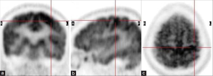
Coronal (a), sagittal (b) and axial (c) positron emission tomography images show increased radiotracer accumulation in the left motor cortex (cross-hairs)

Coronal (a), sagittal (b) and axial (c) positron emission tomography images show increased radiotracer accumulation in the left motor cortex (cross-hairs)