
Figure 11.
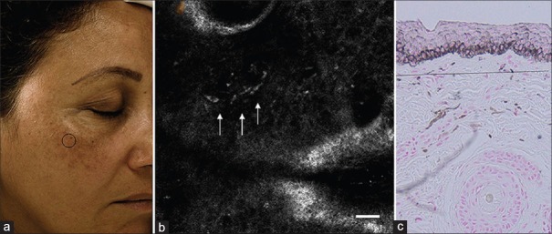
Confocal microscopy image of melasma showing dermal pigmentation: (a) Clinical picture of melasma. (b) Confocal microscopy showing bright plump cells in dermis. (c) Histopathology picture showing melanophages in the dermis (source acknowledged: Kang HY, Bahadoran P, Ortonne JP. Reflectance confocal microscopy for pigmentary disorders. Exp Dermatol 2010;19:233-9)