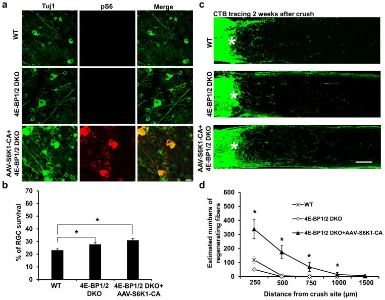
Figure 2. 4E-BP1/2 deletion increases RGC survival but has no effect on axon regeneration.
(a) Confocal images of flat-mounted retinas showing surviving Tuj1 positive RGCs and p-S6 immunostaining, 2 weeks after ON crush. Scale bar, 20 μm. (b) Quantification of surviving RGCs, represented as percentage of Tuj1 positive RGCs in the injured eye, compared to the intact contralateral eye. *: p<0.05. Data are presented as means ± s.e.m, n=8–12. (c) Confocal images of ON longitudinal sections showing regenerating fibers labeled with CTB 2 weeks after ON crush. *: crush site. Scale bar, 100 μm. (d) Quantification of regenerating fibers at different distances distal to the lesion site. *: p<0.05. Data are presented as means ± s.e.m, n=8–12.

