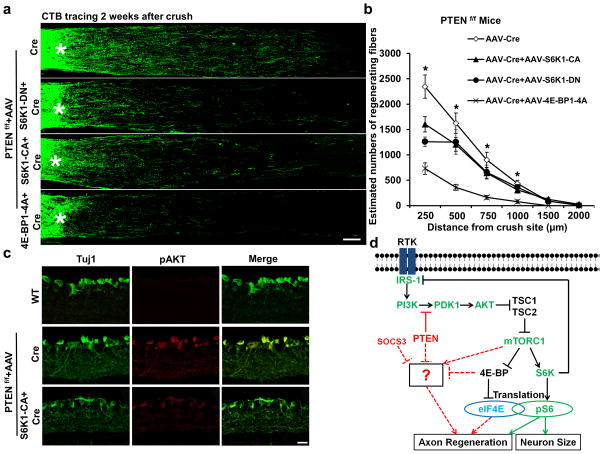
Figure 4. Both S6K1 and 4E-BP are involved in PTEN deletion-induced axon regeneration.
(a) Confocal images of ON longitudinal sections showing regenerating fibers labeled with CTB, 2 weeks after ON crush. *: crush site. Scale bar, 100 μm. (b) Quantification of regenerating fibers at different distances distal to the lesion site. *: p<0.05. Data are presented as means ± s.e.m, n=8–12. (c) Confocal images of retina cross-sections immunostained with Tuj1 and pAkt antibodies, 2 weeks after AAV injection. Scale bar, 20 μm. (d) Schematic summary of the PTEN/mTORC1 signaling pathways in CNS axon regeneration. The dashed lines represent the possible crosstalk among these pathways. The question mark represents unknown effectors downstream of these pathways that contribute to CNS axon regeneration.

