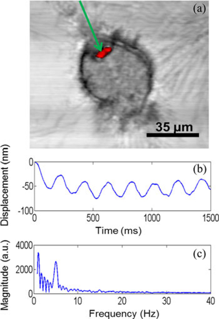
Fig. 4.
Coregistered multimodal imaging and MM-OCE. (a) Simultaneously acquired and coregistered OCM/MPM images of a mouse macrophage that has phagocytosed two fluorescent microspheres (Bangs Labs). The OCM image data are shown in gray-scale, while the two-photon excited fluorescence MPM image data are shown in red. The location of the optical beam for recording MM-OCE displacements is indicated by the green arrow. (b) Plot of sinusoidal axial displacement of the microspheres as calculated from phase data. (c) Frequency spectrum of the magnetomotive signal obtained by taking the Fourier transform of the displacement signal during 5-Hz modulation by the external magnetic field.

