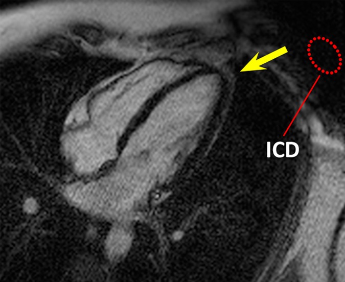
Figure 1c:

HLA LGE MR images in a healthy volunteer, acquired 15 minutes after gadolinium chelate injection. Dotted red ellipse = approximate location of ICD. (a) Image obtained by using the conventional LGE sequence, without the ICD present. (b) Image obtained by using the conventional LGE sequence, with the ICD attached near the shoulder. Hyperintensity artifacts were formed at the apex (arrow). (c) Image obtained by using the proposed wideband LGE sequence, with the ICD attached near the shoulder. Hyperintensity artifact was no longer present.
