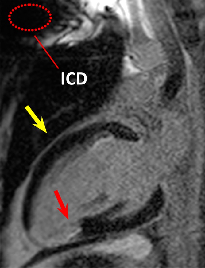
Figure 2b:

LGE images in the two-chamber vertical long-axis plane in patient 1. Dotted red ellipse = approximate location of ICD. (a) Image obtained by using the conventional LGE sequence. Hyperintensity artifact was produced in the ventricular wall (yellow arrow). (b) Image obtained by using the modified wideband LGE sequence. Hyperintensity artifact has been completely resolved (yellow arrow). In this patient, scarring and ventricular wall thinning was present near the posterior LV wall (red arrows), which is remote from the ICD.
