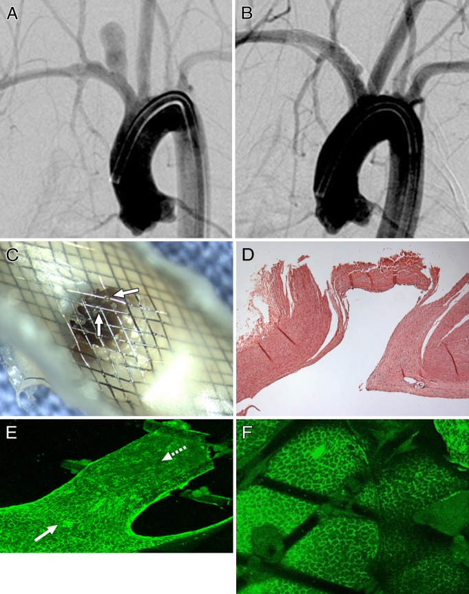
Figure 2:
A, DSA image obtained immediately before flow diverter implantation shows patent aneurysm cavity. B, DSA image obtained 8 weeks after flow diverter implantation shows near-complete occlusion. C, Gross image shows scattered tissue islands over neck (arrows). D, Photomicrograph of histopathologic slice through midportion of neck (hematoxylin-eosin stain; original magnification, ×100) shows neck remnant with endothelialized organized thrombus deep to neck. Tissue seen on C covering the neck was dislodged during processing, so no tissue is present over the neck on this slice. E, Immunostained confocal microscopic image of a tissue island noted in C (CD31 stain; original magnification, ×20). Note confluent coverage with CD31-positive endothelial cells along more peripherally located struts (solid arrow) in direct contiguity with parent artery, with a well-demarcated interface between CD31-positive and CD31-negative (dashed arrow) cells covering more centrally located struts. F, Confocal microscopic image of center of neck (CD31 stain; original magnification, ×20) shows, in foreground, CD31-negative cells attached to intersections of struts and corresponding to tissue islands on C and, in background, deep to struts, confluent endothelial cells as on D.

